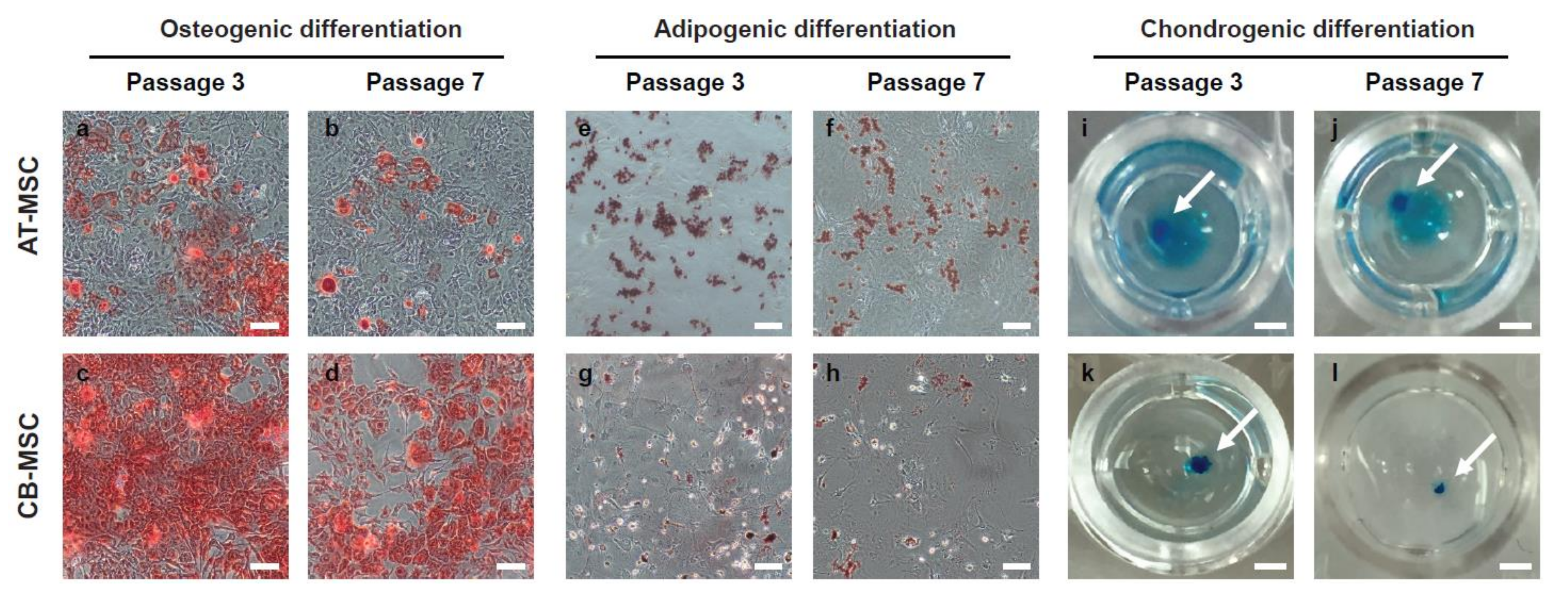
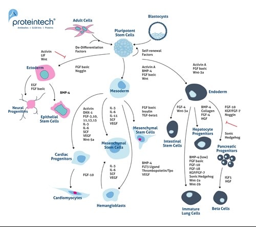
Mesenchymal stem cells mscs have the capacity for multi lineage differentiation giving rise to a variety of mesenchymal phenotypes such as osteoblasts bone adipocytes fat and chondrocytes cartilage.
Flow cytometry protocol mesenchymal stem cells.
Optimization of a flow cytometry based protocol for detection and phenotypic characterization of multipotent mesenchymal stromal cells from human bone marrow.
Springer nature is developing a new tool to find and evaluate protocols.
This unit describes protocols for isolating subpopulations of extracellular vesicles evs purified from human adipose tissue derived mesenchymal stromal cells by density gradient centrifugation and for characterizing them by flow cytometry fcm.
Dental pulp mesenchymal stem stromal cells flow cytometry this is a preview of subscription content log in to check access.
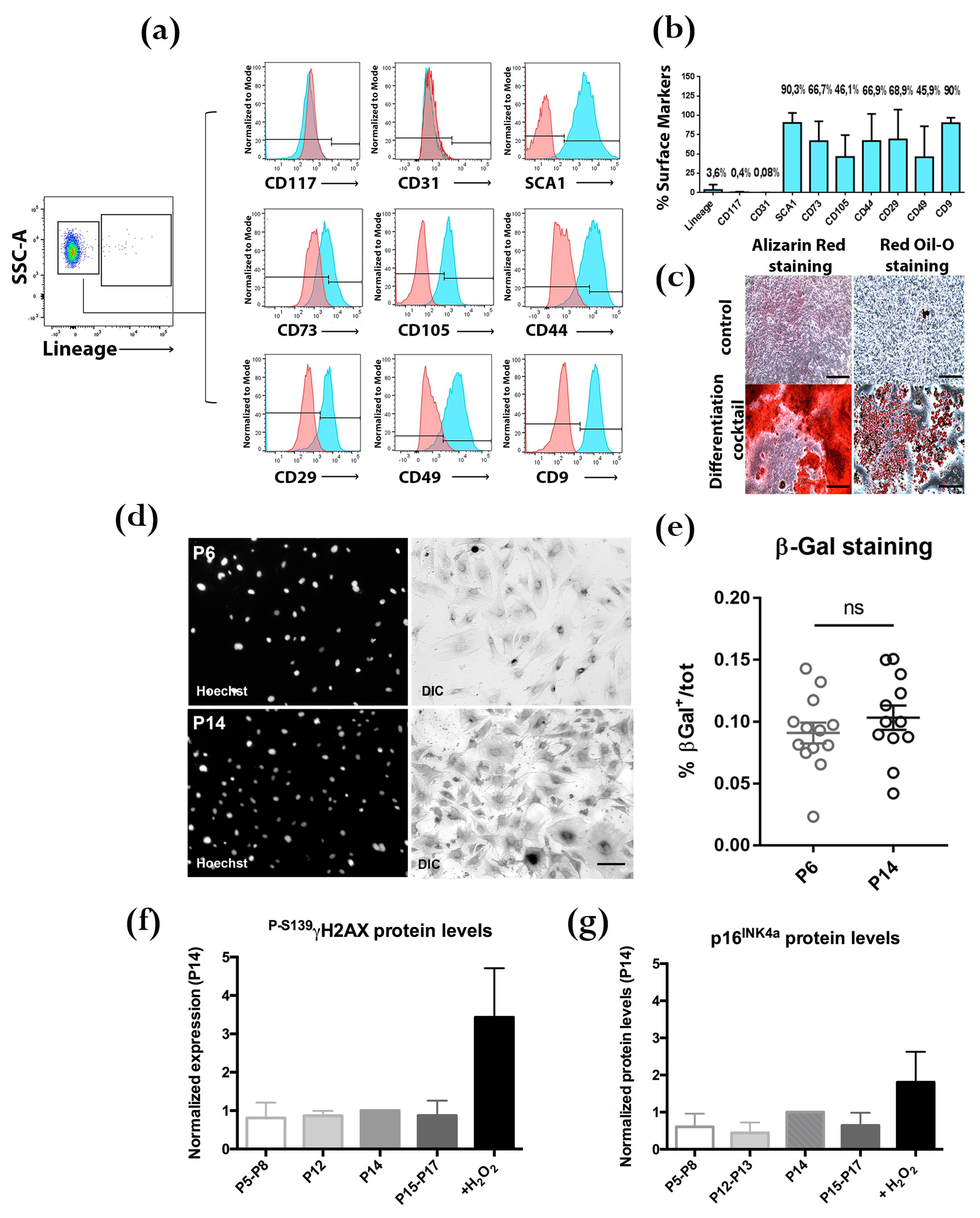
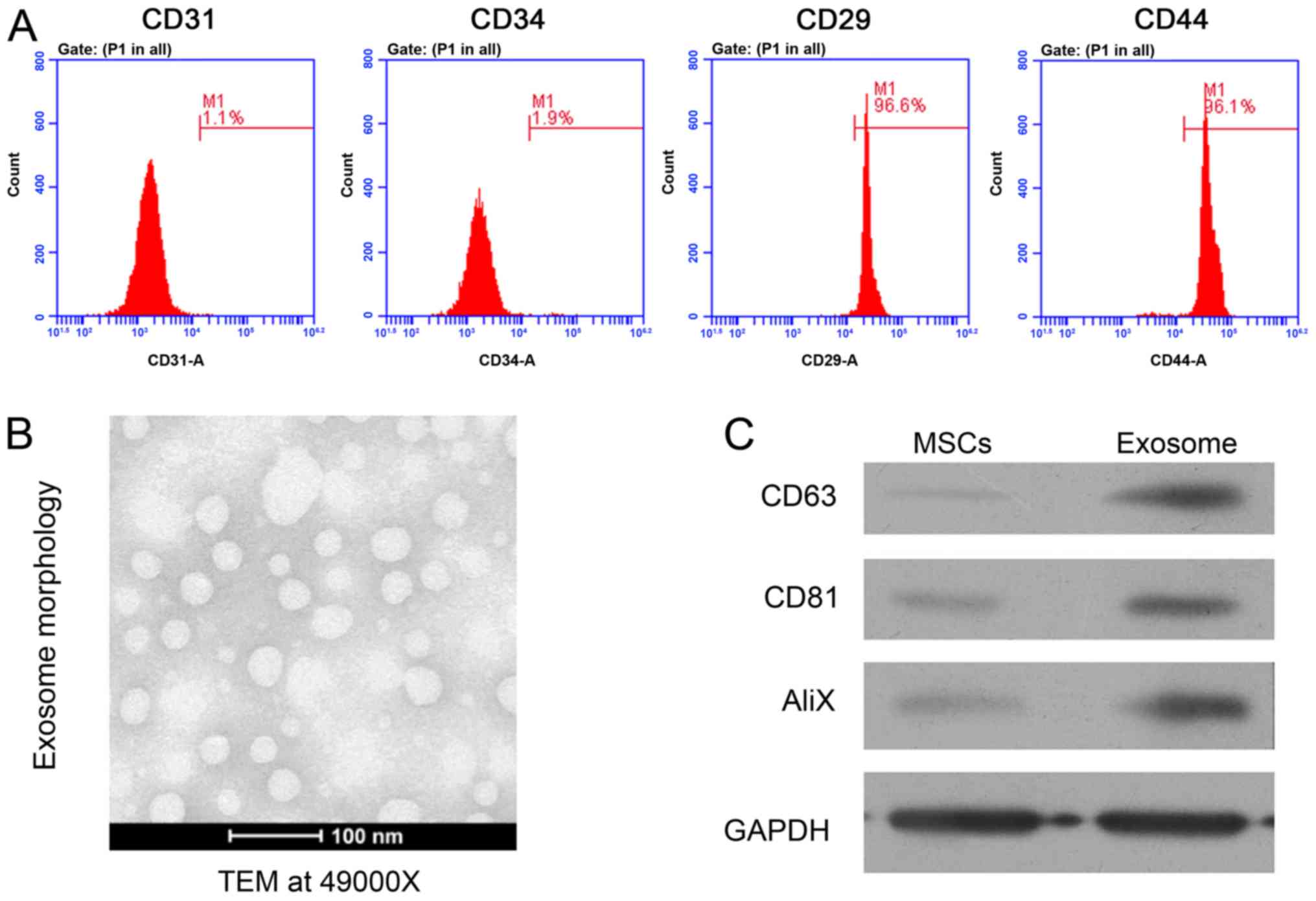
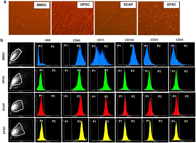
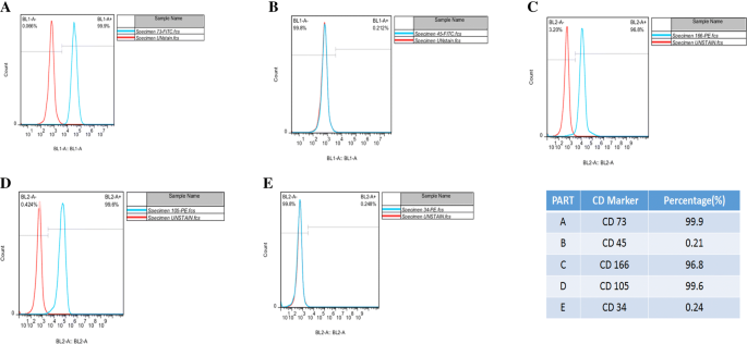
Undifferentiated a and osteocyte differentiated b human mesenchymal stem cells were stained with the indicated antibodies filled histograms or the corresponding isotype control open histograms using the human mesenchymal stromal cell multi color flow cytometry kit catalog fmc002.
Human mesenchymal stem cell flow cytometry kit summary description this product is designed for the flow cytometric analysis of human mscs using four fluorochrome conjugated antibodies.
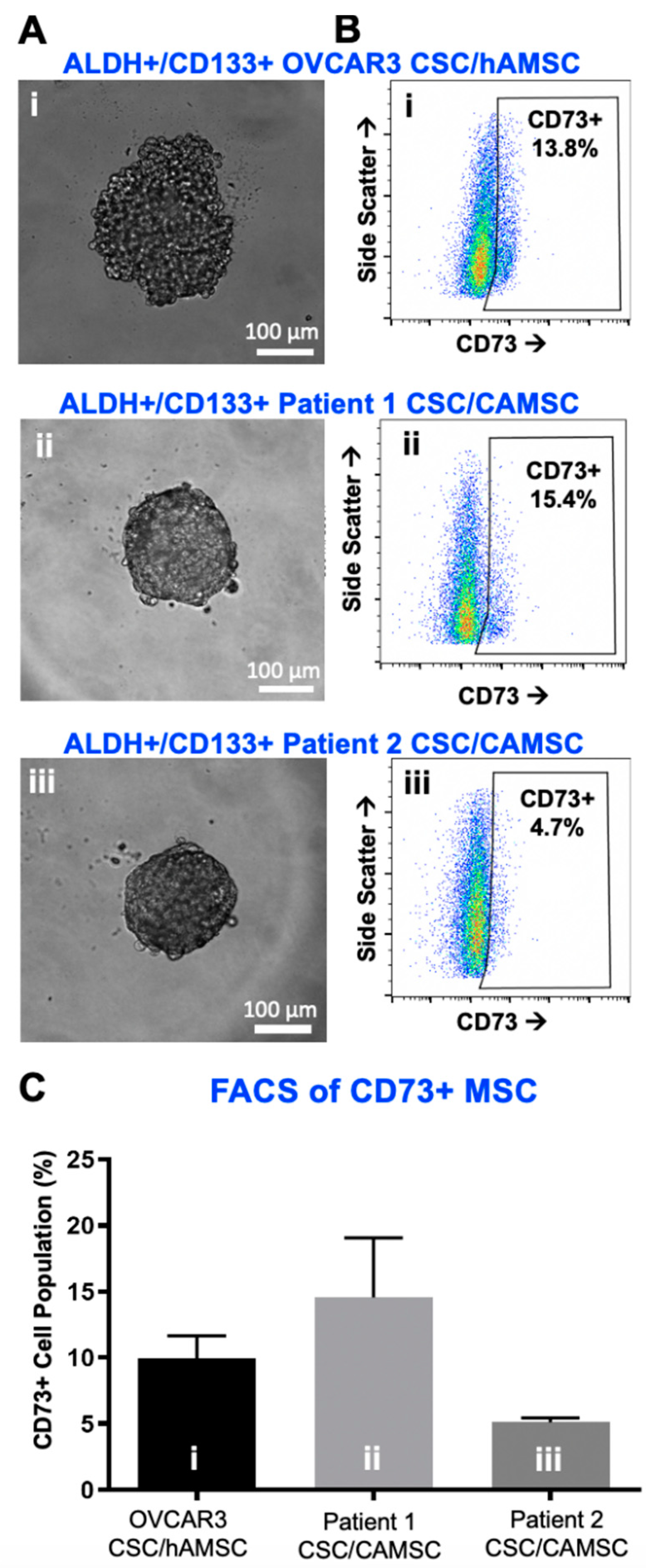
For the simultaneous detection of mesenchymal stem cell markers by flow cytometry.
Cytometry b clin cytom.
We show that flow cytometry an.
Jones ea 1 english a kinsey se straszynski l emery p ponchel f mcgonagle d.
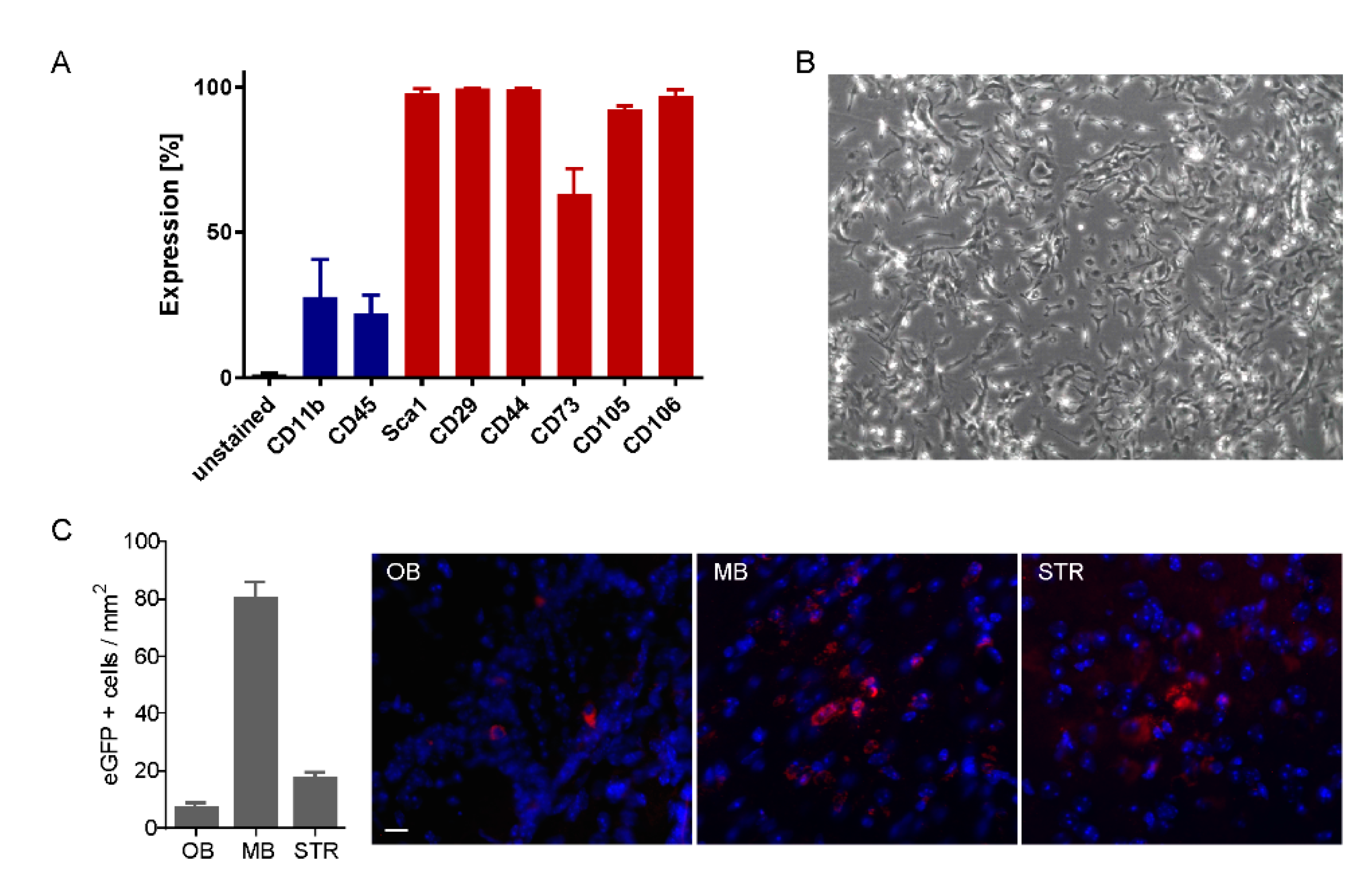
Mesenchymal stem cells mscs are particularly relevant for therapy due to their simplicity of isolation.
Stem cell therapy holds immense promise of delivering the next generation of future medical breakthroughs.
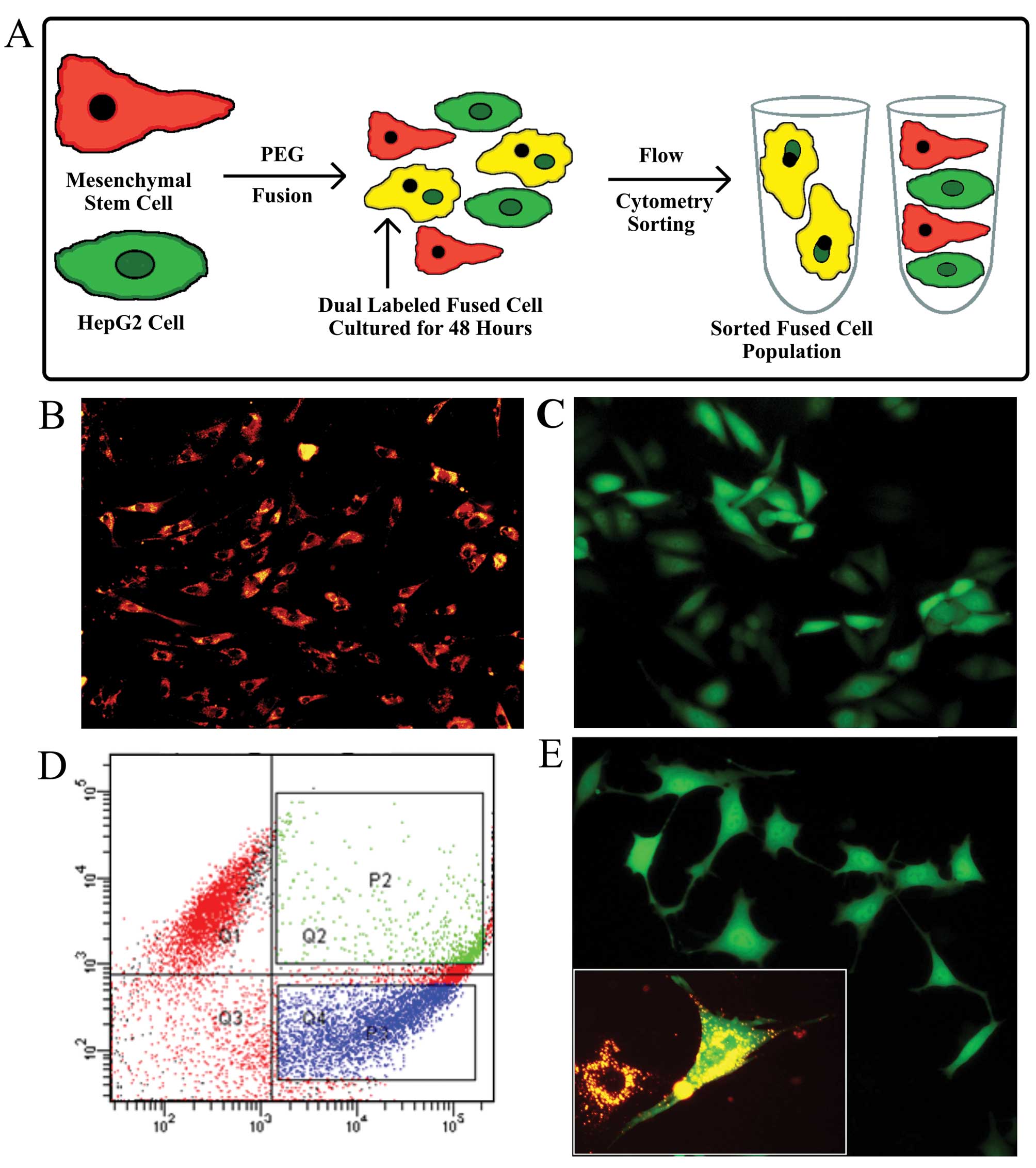
Here we demonstrate the advantages of hyper based ratiometric flow cytometry assay for h2o2 by using k562 and human mesenchymal stem cell lines expressing hyper.
Cell subculture was performed from confluent cultures around 3 days and expanded after trypsinization of the cells.
Mscs grow in monolayer adhered to the surface of the culture dish.
The selection and expansion of the mesenchymal stem cells were performed by successive repackage up to the 10th culture pass.
Multi color flow cytometry simultaneously detected.
